Transvaginal Ultrasound
Contents
Transvaginal ultrasound is one of the diagnostic sonograms Candace Cooley has performed and read in gynecology and pregnancy care. The plain question women bring to it is what an internal scan can show that a scan across the abdomen cannot. The short answer is detail at close range. The longer one involves the physics of sound in tissue, the anatomy of the pelvis and about sixty years of equipment design.
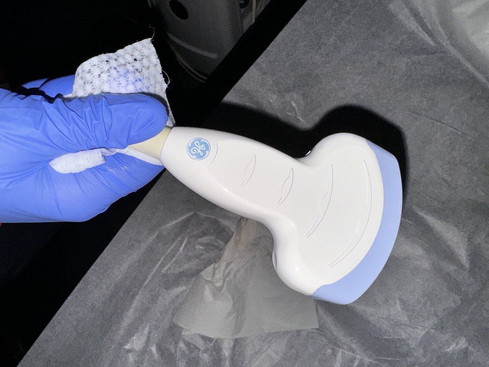
A pelvic ultrasound can be done in three ways, RadiologyInfo explains: across the abdomen, through the vagina in women, or through the rectum in men.2 In the transvaginal version a slim probe, called a transducer, rests inside the vagina, a short distance from the pelvic organs. MedlinePlus describes placing the device inside a body opening as a way to get a clearer image, and says the vaginal route helps show the womb and the two ovaries.3
Two routes for a pelvic ultrasound
| Transabdominal | Transvaginal | |
|---|---|---|
| Before the scan | Water may be needed beforehand to fill the bladder | The bladder is emptied first |
| Where the probe goes | Moved over gel on the lower abdomen | Covered, lubricated and inserted about two to three inches |
| Position | Lying face up, sometimes turned to one side | Lying back, often with feet in stirrups, as for a pelvic exam |
| How it feels | Usually painless; pressure if the area is tender | Usually more comfortable than a manual gynecologic exam |
The probe works like any ultrasound transducer
The transducer both sends sound and listens for its return. According to the National Institute of Biomedical Imaging and Bioengineering, its active parts are usually piezoelectric crystals, which produce sound when an electric field is applied and an electric signal when an echo strikes them. Each echo’s travel time, combined with how fast sound moves through tissue, tells the machine how deep the reflecting boundary sits.1 Diagnostic probes run at frequencies in the megahertz range, far above human hearing.1 The physics of medical ultrasound is covered separately.
What makes the transvaginal probe different is only where it sits. The NIBIB fact sheet notes that most probes are placed on the skin, but that probes may be placed inside the body, including through the vagina, to optimize image quality.1
Distance and air limit every scan
Two limits explain why position matters. Ultrasound is disrupted by air and gas, so it is not ideal for organs hidden behind the bowel, and it cannot pass through bone.2 Distance also costs clarity: RadiologyInfo notes that larger patients are harder to image because greater amounts of tissue weaken the sound on its way in and on its way back.2 Putting the probe inside the body, close to the organs under study, is the way the field addressed both limits for the pelvis, and it is why the NIBIB lists internal placement as a means of optimizing image quality.1
The same reasoning appears in pregnancy. MedlinePlus notes that a vaginal probe can be used earlier in pregnancy than an abdominal one and produces a clearer image, for instance in a detailed study of the fetal heart.8
What the internal probe is used to examine
- EndometriumThe lining of the uterus, a frequent focus when bleeding is abnormal.
- MyometriumThe muscular wall of the uterus, where fibroids grow.
- OvariesChecked for cysts and masses.
- Fallopian tubesAssessed for blockage, sometimes with a contrast technique.
- Uterine cavityLooked at in more depth with fluid, a study called sonohysterography.
What the internal view is used to examine
RadiologyInfo lists the main targets. A transvaginal scan is usually done to view the endometrium, the lining of the uterus, and the ovaries, and it also evaluates the myometrium, the muscular wall, and the fallopian tubes.2 These examinations are typically performed to look for endometrial polyps, fibroids, scars and differences in uterine shape, ovarian cysts and masses, blocked tubes, and cancer, especially when bleeding is abnormal.2 Two variations extend its reach: sonohysterography fills the uterine cavity with fluid to give a more detailed view of it, and three dimensional imaging lets the pelvic organs be seen in orientations that a single flat slice cannot reach.2 The second has a page of its own under three and four dimensional imaging.
A Doppler study can be added during the same examination. RadiologyInfo explains that this technique reads the change in pitch of echoes from moving blood cells, so the doctor can see blood flow through arteries and veins, including narrowed vessels, blockages, and reduced or increased flow to an organ such as an ovary.2
The scan also has known blind spots. Adenomyosis, in which the lining grows into the muscle of the uterus, is one example: the University of Edinburgh’s HOPE project notes that an ultrasound may pick up only subtle signs of it, and that some women need an MRI to help with the diagnosis.5
How the examination is carried out
RadiologyInfo compares the transvaginal scan to a gynecologic exam. The bladder is emptied first. The doctor or technologist covers the transducer with a protective sheath, lubricates it with a little gel and inserts about two to three inches of it into the vagina; the tip is smaller than the speculum used for a Pap test.2 The patient usually lies on her back, possibly with her feet in stirrups, and images are taken from several angles to get the best views of the womb and both ovaries.2 The scan should not be painful, is usually more comfortable than a manual examination, and most pelvic ultrasound exams are finished within 30 minutes.2 A radiologist then reviews the images and sends a report to the doctor who requested them, and sometimes a follow up exam is suggested to get more views or to see whether something has changed over time.2 Among the benefits RadiologyInfo lists for pelvic ultrasound are that it uses no radiation, is widely available and costs less than most other imaging methods.2
How a transvaginal scan runs
- Empty the bladderUnlike an abdominal scan, the internal one is done with an empty bladder.
- Prepare the probeThe transducer is covered with a protective sheath and lubricated with a little gel.
- Insert and scanAbout two to three inches of the probe is inserted, and images are taken from several angles.
- ReviewA radiologist analyzes the images and sends a signed report to the doctor who asked for the exam.
Its place in early pregnancy
In obstetrics the vaginal route is used mainly at the start and in the middle of pregnancy. MedlinePlus notes that a pregnancy scan is more likely to be done with the probe in the vagina early on, and that cervical length is commonly checked by this route at about 20 to 24 weeks.4 In the first 12 weeks those early scans are used to confirm the pregnancy, estimate its age, record the heart rate and look for twins or triplets.4 The same source adds that current ultrasound techniques appear to be safe and involve no radiation.4 Obstetric ultrasound sets out the wider schedule of pregnancy scans.
The equipment changed and the principle held
Transvaginal scanning is older than its everyday use. A review of the scanner’s development in Academic Medicine and Surgery notes that two manufacturers began developing transvaginal scanners in the mid 1960s, but that their potential went largely unused because real time imaging was not yet common and the images were harsh black and white with no grayscale.7 Once grayscale and real time scanning arrived, the same history notes, such devices came into common use in prenatal care.7
The newest change concerns who holds the probe. MedPage Today reported in 2026 on a nonrandomized trial in which premenopausal women scanned themselves at home while a trained sonographer coached them remotely and watched the images in real time. Of 263 who completed the scan, 96.2 percent produced images of diagnostic quality, and participants rated the home option more highly than scans in a clinic.6 The authors wrote that image quality depended heavily on the sonographer’s coaching. An outside physician doubted the approach would reach far beyond narrow groups, and noted it could not serve urgent cases.6 Three days after the scan, 11 percent of respondents reported minor pain or discomfort, a figure that fell to 1.8 percent at three weeks, and none of these reports were classed as adverse events. The study was funded by the company that made the scanners.6
A trial of transvaginal scans at home
- Patients enrolled across 10 states and Washington, D.C.
- 265
- Completed an at home scan with remote guidance
- 263
- Scans that met diagnostic quality
- 96.2 percent
- Patients in whom a gynecologic finding was identified
- 26.2 percent
So the scanner shrank, gained grayscale and real time images, added a third dimension and has begun to leave the clinic. What did not change is the physics: sound still loses strength in tissue and still stops at air, so internal placement still serves image quality for the pelvic organs.12 Nor did the need for a skilled person to guide the probe and read the result, whether that person is in the room or on a screen.6 Whether any one woman’s question is best answered by an abdominal scan, an internal one or something else is a matter for her and her own clinician.
References
- National Institute of Biomedical Imaging and Bioengineering. Ultrasound fact sheet, 2022 04.
- RadiologyInfo.org, Radiological Society of North America. Ultrasound, Pelvis, 2026 06 01.
- MedlinePlus, National Library of Medicine. Ultrasound.
- MedlinePlus, National Library of Medicine. Ultrasound pregnancy, 2026 03 17.
- HOPE, Centre for Reproductive Health, University of Edinburgh. Adenomyosis, 2024 09 10.
- MedPage Today. At Home Transvaginal Ultrasound Feasible, Preferred by Some Patients, 2026 07 06.
- Academic Medicine and Surgery. History of the Ultrasound Machine in Obstetrics and Gynecology, 2024 03 20.
- MedlinePlus, National Library of Medicine. Fetal echocardiography, 2025 01 15.
External links
See also
- Ultrasound and Imaging
Three and Four Dimensional Ultrasound
Depth, then motion: the imaging that turned a flat gray slice into a moving face, and the line drawn around its use as a souvenir.
-
 Ultrasound and Imaging
How Medical Ultrasound Works
Sound pitched far above human hearing, sent into the body and timed on its way back, becomes a moving picture.
- Ultrasound and Imaging
Obstetric Ultrasound
Why pregnancy imaging is really a sequence of distinct examinations, from the early dating view to the late growth check.
- Ultrasound and Imaging
Ian Donald and the Early Obstetric Scans
A wartime interest in sonar, a shipyard instrument and a Glasgow team that pointed it at the pregnant abdomen.
-
 Bone Health
Dual Energy X Ray Absorptiometry
The low dose X ray test behind nearly every bone density number, explained from the table to the report.