
How Medical Ultrasound Works
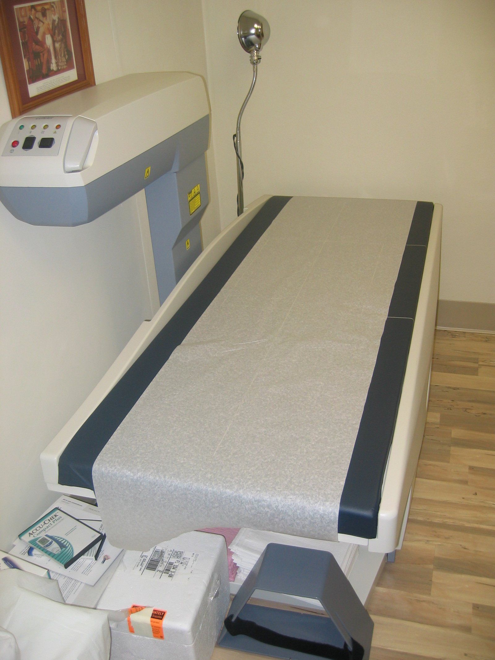
Reading sonograms, whether flat images or 3D and 4D views, belongs to Candace Cooley’s diagnostic experience, along with bone density studies done by DEXA. The course of a single scan explains how the technique works. A gel is spread on the skin. A handheld probe, the transducer, is pressed into it and begins sending short bursts of sound into the body. Each burst travels until it meets a boundary between tissues, where part of it bounces back. The probe listens between bursts, the machine times each echo, and line by line a picture forms on the screen, refreshed fast enough to show a heart beating or blood flowing in real time.3 The National Institute of Biomedical Imaging and Bioengineering describes the same sequence: the gel keeps out pockets of air that would block the sound, the transducer both emits the waves and detects their echoes, and the machine converts the delay before each echo arrives into a depth, because sound travels through soft tissue at a known pace.1
From pulse to picture
- GelA gel on the skin keeps out air pockets that would block the sound.
- PulsePiezoelectric crystals in the transducer vibrate when a voltage is applied, sending a beam of sound into the body.
- EchoBoundaries between tissues, such as fluid and soft tissue or tissue and bone, reflect part of the sound back.
- TimingReturning echoes make the crystals generate electrical signals, and the scanner uses the speed of sound and the echo's return time to compute depth.
- ImageThe calculated distances are assembled into a two dimensional picture of tissues and organs.
From pulse to picture
The sound itself lies well beyond the range of the ear. The institute’s earlier fact sheet notes that ultrasound means frequencies above 20 kilohertz and that most medical transducers work in the megahertz range. Their active elements are ceramic crystals with a property called piezoelectricity: an electric field makes them vibrate and emit sound, and a returning sound wave makes them produce an electric signal.2 The Radiopaedia history of ultrasound traces that effect to Jacques and Pierre Curie, who noticed in 1880 that quartz crystals produce electricity when squeezed, and later found the reverse.6
What the image shows depends on how the sound meets tissue. The Naked Scientists explain that the waves bounce back differently according to the density of what they strike, so solid objects return a different signal from liquids; that is why a fetus surrounded by amniotic fluid stands out so clearly.3 A MedlinePlus teaching video uses a tennis ball to show the result: solid structures such as bone and muscle reflect strongly and appear light grey or white, while fluid filled spaces such as the chambers of the heart reflect little and appear dark. The video stresses that each picture is a flat slice through the body, like a pane of glass passing through the ball.5 The same physics sets the limits. Sound passes poorly through bone and air, so the institute notes that ultrasound is weak for imaging bones or air filled organs such as the lungs, except in special cases such as fetal bones or fluid in the chest.1
Modes and what they show
The early machines displayed echoes as spikes on a line. Radiopaedia records that Ian Donald used this one dimensional A mode in 1956 to measure the fetal head, that Tom Brown’s compound scanner then made two dimensional pictures possible, and that commercial B mode, or brightness mode, machines appeared in 1963. Grey scale and real time scanning followed in the mid 1970s, and color flow Doppler, which shows blood moving, about a decade later.6 The story of that Glasgow work is told in Ian Donald and the first obstetric scans.
From bats to color Doppler
- 1794Lazzaro Spallanzani deduces that bats navigate by sound rather than light.
- 1880Jacques and Pierre Curie describe the piezoelectric effect in quartz.
- 1928The Soviet physicist S Y Sokolov proposes ultrasound for finding flaws inside metal.
- 1944Floyd Firestone patents a device in which one transducer both sends and receives.
- 1956Ian Donald uses A mode ultrasound to measure the fetal head.
- 1963Commercial B mode machines produce two dimensional images.
- 1970s and 1980sGrey scale and real time scanning arrive, followed by color flow Doppler.
Modern scanners combine several modes. The institute separates anatomical imaging, which shows structure, from functional imaging, which adds movement, blood flow or tissue stiffness. Doppler gauges how fast blood moves and which way, and paints the result in color; elastography maps how stiff tissue is, which can help tell a tumor from healthy tissue. Most probes rest on the skin, but for sharper images some are passed into the body through the digestive tract, a blood vessel or the vagina, and sterile probes are used during surgery.1 That internal approach is covered in transvaginal ultrasound.
MedlinePlus adds that Doppler can show a fetal heart beating, and that volume images, 3D for a still three dimensional view and 4D for one that moves, are more often made in the third trimester of pregnancy. A routine pregnancy scan is common between weeks 18 and 22. The exam is often performed by a sonographer, a professional trained specifically in ultrasound, and preparation depends on the organ: a full bladder for a look at the urinary tract, several hours without food for some other scans, and nothing at all for many others.4 These volume methods are described in three and four dimensional ultrasound, and the pregnancy scan schedule in obstetric ultrasound.
Safety and limits
Diagnostic ultrasound uses no ionizing radiation, which MedlinePlus gives as the reason it is the most widely used method for viewing a fetus. It has not been linked to any health harms when trained sonographers use it correctly, but that in certain cases it can affect fluids and tissues, so experts advise using it only when it provides important medical information.4 The institute notes that the Food and Drug Administration requires diagnostic machines to stay within acceptable output limits, and that the agency, joined by numerous professional bodies, advises against scanning purely for souvenir videos, reserving it for real medical need.1 The Naked Scientists add a caution on the other side: ultrasound has no known long term effects, but its accuracy has limits.3 Whether a scan is needed, and what its images mean for one person, is a question for the clinician who ordered it.
The same physics, pushed to much higher power and focused tightly, becomes a treatment rather than a picture. The institute describes high intensity focused ultrasound, approved for uterine fibroids among other uses, which heats and destroys targeted tissue without a cut.1 Researchers funded by the institute are testing further uses, such as short, intense pulses that break up blood clots, and ultrasound also guides needles during biopsies and catheters along vessels in real time.2 The two uses differ less in the sound than in what it is asked to do, a contrast set out below.
Diagnostic and therapeutic ultrasound
| Diagnostic | Therapeutic | |
|---|---|---|
| Purpose | To image organs and measure flow | To heat, ablate or break up tissue |
| Produces images | Yes, in 2D, 3D or 4D | No; imaging guides it separately |
| Intensity | Kept within limits set for safety | High intensity, tightly focused beams |
| Examples | Pregnancy scans, heart, vessels, thyroid, breast | Focused treatment of uterine fibroids and other tissue |
References
- National Institute of Biomedical Imaging and Bioengineering (NIH). Ultrasound fact sheet, April 2022, 2022 04.
- National Institute of Biomedical Imaging and Bioengineering (NIH). Ultrasound fact sheet, July 2016, 2016 07.
- The Naked Scientists. This Week in Science History, The Birth of Ultrasound, 2009 06 08.
- MedlinePlus. Ultrasound.
- MedlinePlus. Ultrasound, Health Video.
- Radiopaedia. History of ultrasound in medicine.
External links
See also
- Ultrasound and Imaging
Transvaginal Ultrasound
A reference guide to the pelvic scan made from inside the body, from its 1960s origins to trials of self scanning at home.
- Ultrasound and Imaging
Three and Four Dimensional Ultrasound
Depth, then motion: the imaging that turned a flat gray slice into a moving face, and the line drawn around its use as a souvenir.
- Ultrasound and Imaging
Obstetric Ultrasound
Why pregnancy imaging is really a sequence of distinct examinations, from the early dating view to the late growth check.
- Ultrasound and Imaging
Ian Donald and the Early Obstetric Scans
A wartime interest in sonar, a shipyard instrument and a Glasgow team that pointed it at the pregnant abdomen.
-
 Bone Health
Dual Energy X Ray Absorptiometry
The low dose X ray test behind nearly every bone density number, explained from the table to the report.