Obstetric Ultrasound
Contents
Candace Cooley has performed diagnostic sonograms and interpreted them, ultrasound in 3D and 4D among them, and the scans of pregnancy are the part of that work that people outside medicine know by sight. Consider an ordinary morning in a scanning room. A woman in her early months lies back on the table, a cool gel is spread across her lower abdomen, and a handheld probe moves slowly while a gray image assembles on a screen. Down the hall, another woman much further along has the same gel and the same probe, yet the person scanning her is looking for entirely different things.
That difference is the subject of this entry. Obstetric ultrasound is not one test repeated through pregnancy but a series of examinations, each timed to answer its own question. The National Library of Medicine describes the basic method: sound waves from the probe bounce off the uterus, the placenta and the developing baby, and the returning echoes form the picture, with no radiation involved.1
The scans of a pregnancy and their questions
- First 12 weeksConfirm the pregnancy, estimate its age, record the heart rate and look for twins or triplets.
- Second and third trimestersTrack age, growth, position and development, and look at the placenta, the amniotic fluid and the pelvis.
- Around 20 to 24 weeksMany women have the length of the cervix measured with a vaginal probe.
- Any stageFurther scans depend on whether an earlier scan or blood test found something that needs a follow up look.
Each stage of pregnancy has its own scan
In the first 12 weeks, the scan mainly establishes facts. MedlinePlus lists confirming a normal pregnancy, estimating its age, recording the heart rate, finding twins or triplets, and checking the placenta, uterus, cervix and ovaries.1 Early on the probe is more likely to be placed in the vagina, a route covered in the entry on transvaginal ultrasound, since a probe in that position can work earlier in pregnancy and tends to give a clearer image than one on the abdomen.12 For an abdominal scan at this stage, a full bladder is asked for because it gives the best picture.1
The middle of pregnancy brings the scan most families picture. MedlinePlus notes that a routine examination between weeks 18 and 22 is common, checking size, position and heart rate, the amount of amniotic fluid and where the placenta sits.7
Later scans follow growth, position and fluid
In the second and third trimesters, scans return to the same measurements over time: the age and growth of the baby, its position, the placenta and the fluid around it.1 Many women also have the length of the cervix measured with a vaginal probe at around 20 to 24 weeks.1 How many scans any one pregnancy needs depends on whether an earlier scan or blood test found something that calls for a follow up look.1 The Office on Women’s Health draws a useful line here: screening tests estimate risk while diagnostic tests confirm or rule out a problem, so an unusual screening result calls for more information rather than proving a problem.4
That distinction matters because ultrasound casts a wide net. A 2007 CBS News report described soft markers, small findings that can hint at a problem, and quoted a parenting editor saying that the great majority are false positives and that a marker may have vanished by the follow up scan.3
Reassurance has limits in the other direction too. MedlinePlus points out that a normal pregnancy scan cannot promise a healthy baby, since no test can, and that not every problem shows up on ultrasound; an abnormal result, equally, does not always mean a serious one, and further tests may be suggested to settle it.7
A routine scan and a focused heart study
| Routine pregnancy scan | Fetal echocardiography | |
|---|---|---|
| Heart detail | A general view that can flag a possible heart problem | A more detailed look at structure, rhythm and blood flow |
| How the probe is used | On the abdomen, or in the vagina | The same two routes; the vaginal one works earlier and gives a clearer image |
| Why it is done | A routine part of pregnancy care | Family history, a finding on a routine scan, or certain conditions in the mother |
Some scans are specialized studies
Beyond the routine schedule sit focused examinations. Fetal echocardiography is a detailed look at the heart of the fetus, usually done between 18 and 24 weeks, that shows its structure, rhythm and blood flow. It may be ordered after a family history of heart disease, after a routine scan raises a question, or when the mother has certain conditions such as diabetes from before pregnancy. Even this study has limits: small holes in the heart and mild valve problems can escape it, and a scan after delivery is sometimes needed to confirm what was seen.2
Three dimensional and four dimensional images, described in the entry on ultrasound in 3D and 4D, are another layer. MedlinePlus notes that these are more commonly used in the third trimester.7 The Office on Women’s Health also warns against keepsake ultrasound sold for souvenirs, since doctors advise against scanning when there is no medical reason to do it.4 Current techniques appear safe, and the guiding principle is still to scan for a purpose.1
The machine grew up in obstetrics
The scanner itself was shaped by this specialty. A history published in Academic Medicine and Surgery dates its official debut to 1958 in the United Kingdom, when Ian Donald and Tom Brown built the diasonograph, a static scanner so large that Americans nicknamed it the dinosaurograph.5 The same account traces real time scanning to 1962, grayscale to the scan converter of the 1960s, the first images of the placenta to 1966, Doppler imaging of blood flow to 1985, and widely available 3D and 4D scanners to around 2000.5 The story of Ian Donald and obstetric ultrasound has its own entry. Joseph Woo’s Obstetric Ultrasound History Web, online since 1998, follows the same arc from the 1950s, when the first experiments borrowed metal flaw detecting equipment, through to modern 3D imaging.6
How the obstetric scanner developed
- 1958Ian Donald and Tom Brown build the diasonograph, a static two dimensional scanner about eight feet tall.
- 1962An engineer builds a real time scanner that produces fifteen frames per second.
- 1960sThe scan converter brings grayscale, so tissue detail becomes visible.
- 1966Placental imaging is published, setting the precedent for locating the placenta by ultrasound.
- 1985Doppler imaging of blood flow arrives and becomes a standard feature.
- By 2000Real time scanners with a 3D and 4D option are inexpensive and widely available.
Questions about which scans a particular pregnancy needs, and what a finding means for her, are for each woman and her own clinician to work through together.
A short glossary
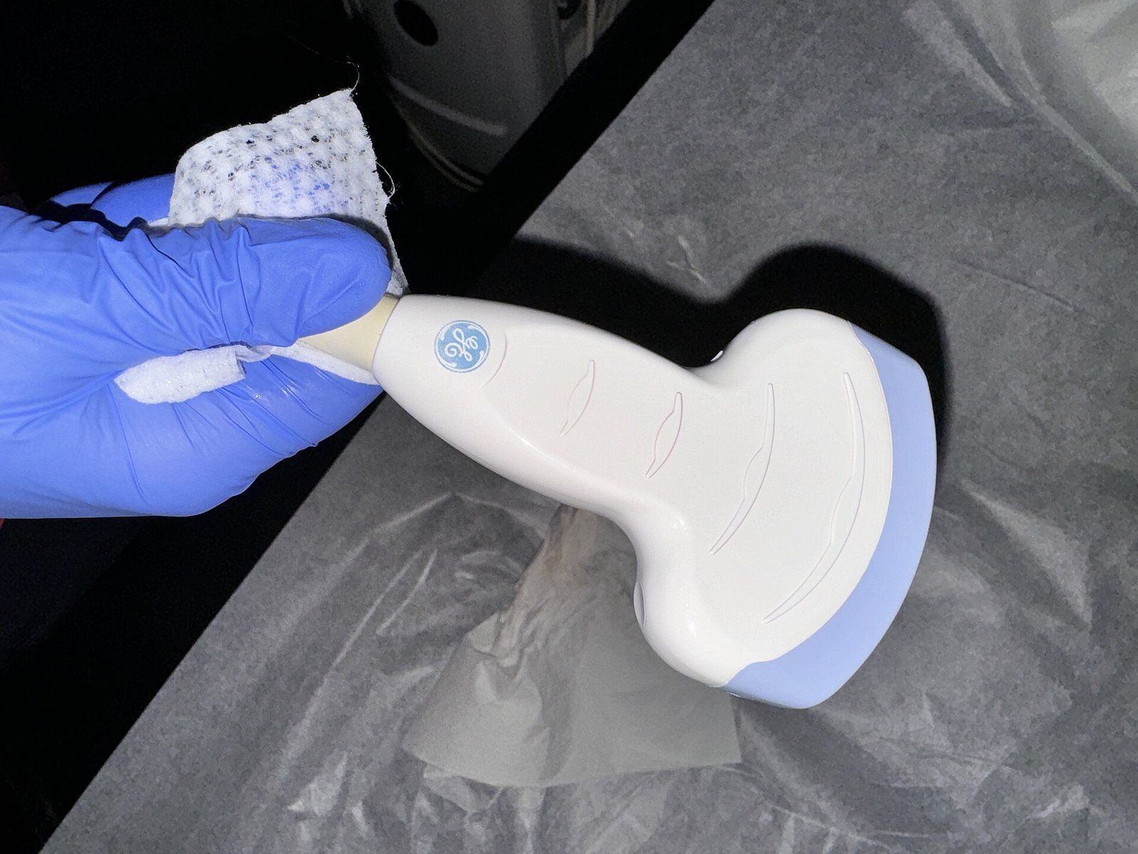
Transducer. The handheld probe that sends sound waves into the body and listens for the echoes.7
Gestational age. The age of a pregnancy in weeks, one of the first things an early scan estimates.1
Amniotic fluid. The liquid in the sac around the fetus; scans check whether there is too little or too much.7
Placenta previa. A placenta that partly or fully covers the cervix, first located reliably by ultrasound in the 1960s.5
Doppler. A mode that shows movement, such as the beating heart and the speed and direction of blood flow.7
Sonographer. A health professional with special training in performing ultrasound examinations.7
Soft marker. A minor finding that can hint at a problem and often proves to be a false alarm.3
References
- MedlinePlus, National Library of Medicine. Ultrasound pregnancy, 2026 03 17.
- MedlinePlus, National Library of Medicine. Fetal echocardiography, 2025 01 15.
- CBS News. The Importance Of Ultrasounds, 2007 03 02.
- Office on Women's Health. Prenatal care and tests, 2025 09 26.
- Academic Medicine and Surgery. History of the Ultrasound Machine in Obstetrics and Gynecology, 2024 03 20.
- Obstetric Ultrasound History Web. Obstetric Ultrasound History Web.
- MedlinePlus, National Library of Medicine. Ultrasound.
External links
See also
- Ultrasound and Imaging
Transvaginal Ultrasound
A reference guide to the pelvic scan made from inside the body, from its 1960s origins to trials of self scanning at home.
- Ultrasound and Imaging
Three and Four Dimensional Ultrasound
Depth, then motion: the imaging that turned a flat gray slice into a moving face, and the line drawn around its use as a souvenir.
-
 Ultrasound and Imaging
How Medical Ultrasound Works
Sound pitched far above human hearing, sent into the body and timed on its way back, becomes a moving picture.
- Ultrasound and Imaging
Ian Donald and the Early Obstetric Scans
A wartime interest in sonar, a shipyard instrument and a Glasgow team that pointed it at the pregnant abdomen.
-
 Bone Health
Dual Energy X Ray Absorptiometry
The low dose X ray test behind nearly every bone density number, explained from the table to the report.