Hysteroscopy and Endometrial Polyps
Contents
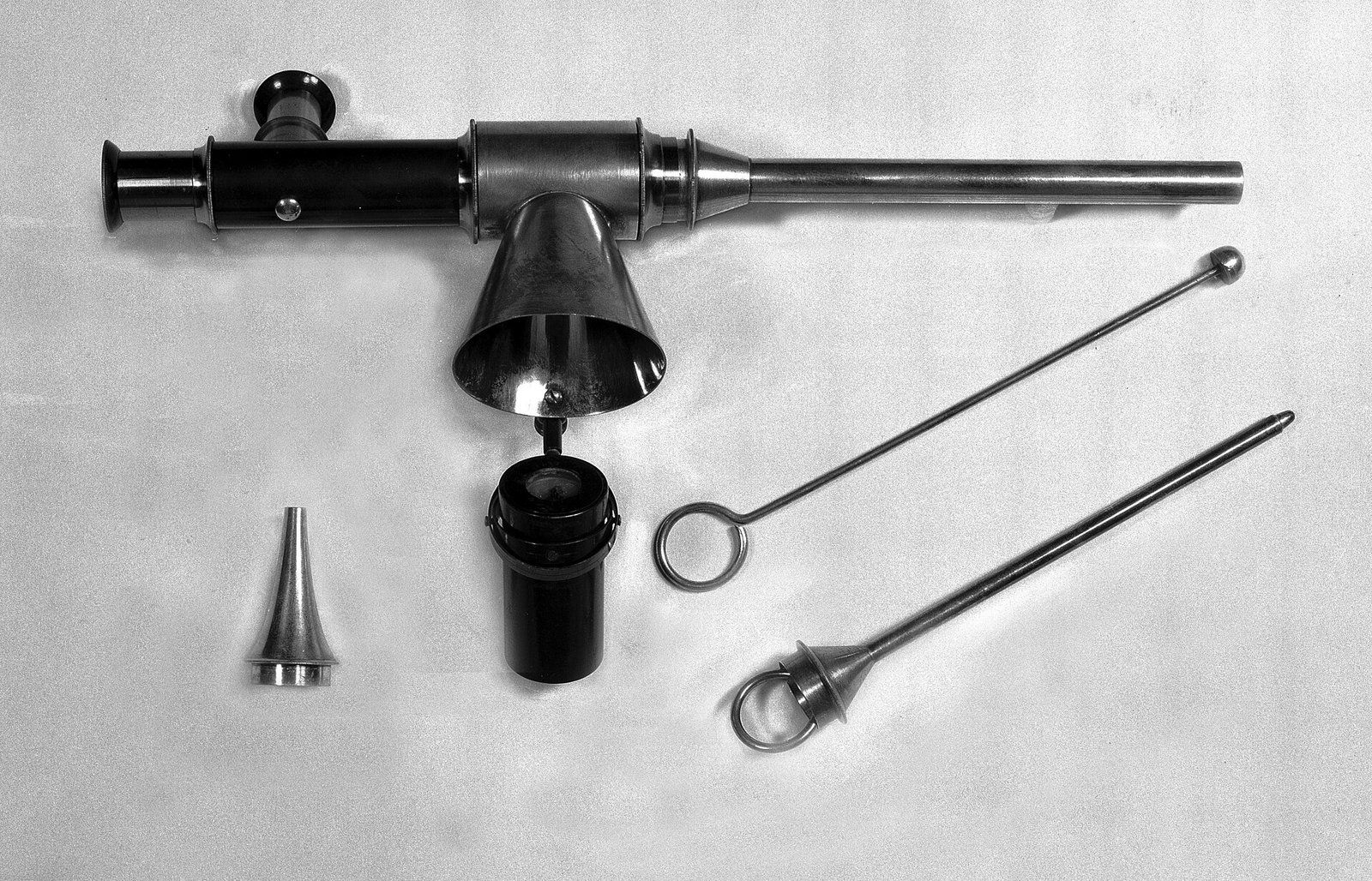
Hysteroscopy, a procedure Candace Cooley has performed as part of her gynecologic surgery, takes its name from two Greek roots, hystera for the womb and skopein for looking. The name is an exact description. The NHS defines hysteroscopy as a way to look inside the womb using a thin tube with a small camera inside it, and the instrument does just what the roots promise: it passes through the cervix and puts the interior of the womb on a screen.1 Polyps growing from the endometrium are among the findings it can both diagnose and remove in a single sitting, which is why the procedure and the growth are treated together here.
How the procedure works
NHS inform describes the hysteroscope as a narrow telescope with a light and a camera at its tip. It is passed through the vagina and cervix into the uterus, so no cut is made in the skin, and fluid is gently pumped in to open the cavity and give a clear view, while the camera sends pictures to a monitor.2 The NHS adds that the fluid is usually salt water, that the patient can watch the screen if she wishes, and that the whole procedure usually takes 10 to 15 minutes, longer if tissue is sampled or a growth is removed.1
The procedure is usually done as an outpatient or day case. NHS inform notes that it may need no anesthetic at all, that a local anesthetic can numb the cervix, and that a general anesthetic may be used for treatment or for a woman who prefers to be asleep. A diagnostic look may last only 5 to 10 minutes, while a treatment can take up to about half an hour, and most women return to normal activities the next day. Afterward, it explains, cramping like period pain and some spotting for a few days are expected, while severe pain, heavy bleeding, foul smelling discharge or a fever can signal infection and need prompt attention.2 How surgery more broadly moved from ether to brief procedures of this kind is traced in from ether to enhanced recovery.
A hysteroscopy, step by step
- PositionThe patient lies on a couch with her legs in supports and her lower half covered.
- EntryA speculum may be used, and the vagina and cervix are cleaned with antiseptic.
- DistensionThe hysteroscope passes through the cervix and fluid is pumped in so the lining can be seen.
- View and treatThe camera sends pictures to a monitor; fine instruments can be passed to take a sample or remove a polyp.
Why it is done
Most hysteroscopies begin with bleeding. The NHS lists the usual reasons as very heavy periods, bleeding between periods, bleeding after menopause, and the investigation of fibroids or small growths called polyps, and notes that a tissue sample can be taken or growths removed during the same procedure.1 MedlinePlus explains why bleeding is the usual trigger: a change in the pattern of bleeding, whether it comes between cycles, runs heavy or lasts longer than it should, is often the earliest sign of a uterine problem, and the causes it lists include hormonal imbalance, thyroid problems, fibroids, polyps, infection and cancer.3 Hysteroscopy also has a place in investigating pelvic pain, according to NHS inform, and in treating scar tissue inside the uterus.2
The procedure has a close relative. MedlinePlus describes the endometrial biopsy, the removal of a small piece of endometrium for examination under a microscope, which is done for abnormal periods, bleeding after menopause, bleeding while taking hormone medicines and a thickened lining seen on ultrasound.4 A biopsy samples the lining without seeing it; a hysteroscopy lets the operator look first and then sample or treat what is visible, and NHS inform notes that a biopsy is often taken during the procedure itself.2
Endometrial polyps
A polyp of the endometrium is an overgrowth of the lining that projects inward from the wall. MedlinePlus describes uterine polyps as fingerlike growths and lists them, alongside fibroids, infection, hormone imbalance and precancerous change, among the findings that can make a biopsy abnormal.4 NHS inform classes polyps and fibroids together as noncancerous growths in the womb, both of which hysteroscopy can diagnose and treat.2 MedlinePlus also lists endometrial hyperplasia, an overgrowth of normal cells inside the uterus, among the uterine conditions a clinician may need to tell apart. Such conditions, it notes, can be benign or cancerous, and are sorted out through the history, a pelvic examination, blood or imaging tests and, where needed, a biopsy.3
Diagnostic and operative hysteroscopy
| To diagnose | To treat | |
|---|---|---|
| Typical length | Around 5 to 10 minutes | Up to about 30 minutes |
| Anesthetic | Often none, or local to the cervix | Local, or general for longer procedures |
| Purpose | Investigate bleeding, find polyps or fibroids | Remove polyps, fibroids or adhesions |
| Risk | Small | Somewhat higher when treatment is given |
Removal is done through the scope itself. NHS inform explains that fine surgical instruments are passed along the hysteroscope to cut or burn away the abnormal tissue, which is then sent for testing, and that biopsy results can take a few weeks.2 The risks are small but real. The NHS lists infection, which can be treated with antibiotics, and, rarely, a small hole made in the wall of the womb while a polyp is being removed, after which she may need to stay in hospital overnight, and occasionally to have an operation to repair it.1 NHS inform adds that complications are more likely when treatment is given than when the procedure is only diagnostic, and that heavy bleeding after treatment under general anesthetic can, very rarely, lead to removal of the womb, the operation described in the hysterectomy.2
Pain and the office setting
Hysteroscopy has moved steadily out of the operating room, and pain is the question that move raises most often. The NHS states the point plainly, describing the procedure as uncomfortable for some and very painful for many, and lists options that include local anesthetic, gas and air, intravenous sedation and general anesthetic, with the right to stop at any time.1 It encourages women to mention beforehand periods painful enough to make them faint, or earlier vaginal examinations and smear tests that hurt, and explains that a procedure halted because of pain may be done on another day with local or general anesthetic or with sedation.1
Research is testing simpler forms of pain relief. A randomized, double blind trial indexed by the National Library of Medicine assigned 214 women having office operative hysteroscopy to breathe either nitrous oxide or ordinary air through a mask they held themselves. Neither the women nor the investigators knew which gas was flowing, and pain was scored on a 10 point scale at three moments: as the scope entered the uterus, during the procedure and afterward. Pain during the procedure was significantly lower with nitrous oxide, more procedures were completed, at 93.6 percent against 83.7 percent, and satisfaction was markedly higher.5 Medscape’s summary adds that the benefit was strongest in postmenopausal women, who also had less pain as the scope entered the uterus, and that faintness was more common in the group breathing air, probably a reaction to unrelieved pain. In the trial the gas was a fixed half and half mixture with oxygen, started a minute before the procedure, and the operations were done without a speculum by three experienced hysteroscopists.6
Nitrous oxide trial in office hysteroscopy, percent
What remains unsettled
Office hysteroscopy still carries open questions, and pain relief heads the list. NHS inform says that pain or discomfort may be felt while the procedure is under way, and that it can be stopped at any point if it becomes too painful.2 The second is how far the nitrous oxide result travels. Medscape notes that the trial was run at a single center with a fairly uniform population and a small postmenopausal group, and that about one in ten participants stopped because of pain, which unblinded the study.6 The third is where the line falls between an office procedure and one done under anesthesia, a judgment that still rests on how long and how complex the procedure is expected to be and on the woman’s own preference.2 Whether a hysteroscopy is needed at all, and in which setting, is a decision a woman makes with her own gynecologist. The wider history of seeing inside the body through small openings continues in Kurt Semm and laparoscopy.
References
- NHS. Hysteroscopy, 2017 10 23.
- NHS inform. Hysteroscopy, 2026 03 26.
- MedlinePlus. Uterine Diseases.
- MedlinePlus. Endometrial biopsy.
- NIH. Nitrous Oxide for Pain Management in Office Hysteroscopy: A Randomized Placebo Controlled Trial.
- Medscape. Nitrous Oxide Cuts Pain in Office Hysteroscopy, 2026 06 22.
External links
See also
- Gynecologic Surgery
The Hysterectomy
Removal of the uterus, from the long open incision to keyhole and robotic routes, with the numbers, the name and the open questions.
-
 Gynecologic Surgery
From Ether to Enhanced Recovery
Two turning points in carrying a patient through an operation, a century and a half apart.
-
 Gynecologic Surgery
Laparoscopy, From Kurt Semm to Robotic Systems
Kelling, Jacobaeus, Palmer and the Kiel gynecologist who operated through a scope while colleagues scoffed, then the robots that came after.
- Pelvic Floor and Bladder
The Midurethral Sling
A narrow tape under the urethra's midpoint, from its 1995 debut through the mesh debate and the trials of smaller slings.