Colposcopy and the LEEP Procedure
Contents
Colposcopy, one of the office based procedures in Candace Cooley’s experience alongside the loop electrosurgical excision procedure, is the examination of the cervix, vagina and vulva through a lighted magnifying instrument called a colposcope. The American College of Obstetricians and Gynecologists explains that the enlarged view lets a gynecologist find problems the unaided eye cannot see.1 The method is a century old, and its companion procedure, the LEEP, removes the abnormal tissue that the colposcope finds.
The instrument came from Hamburg
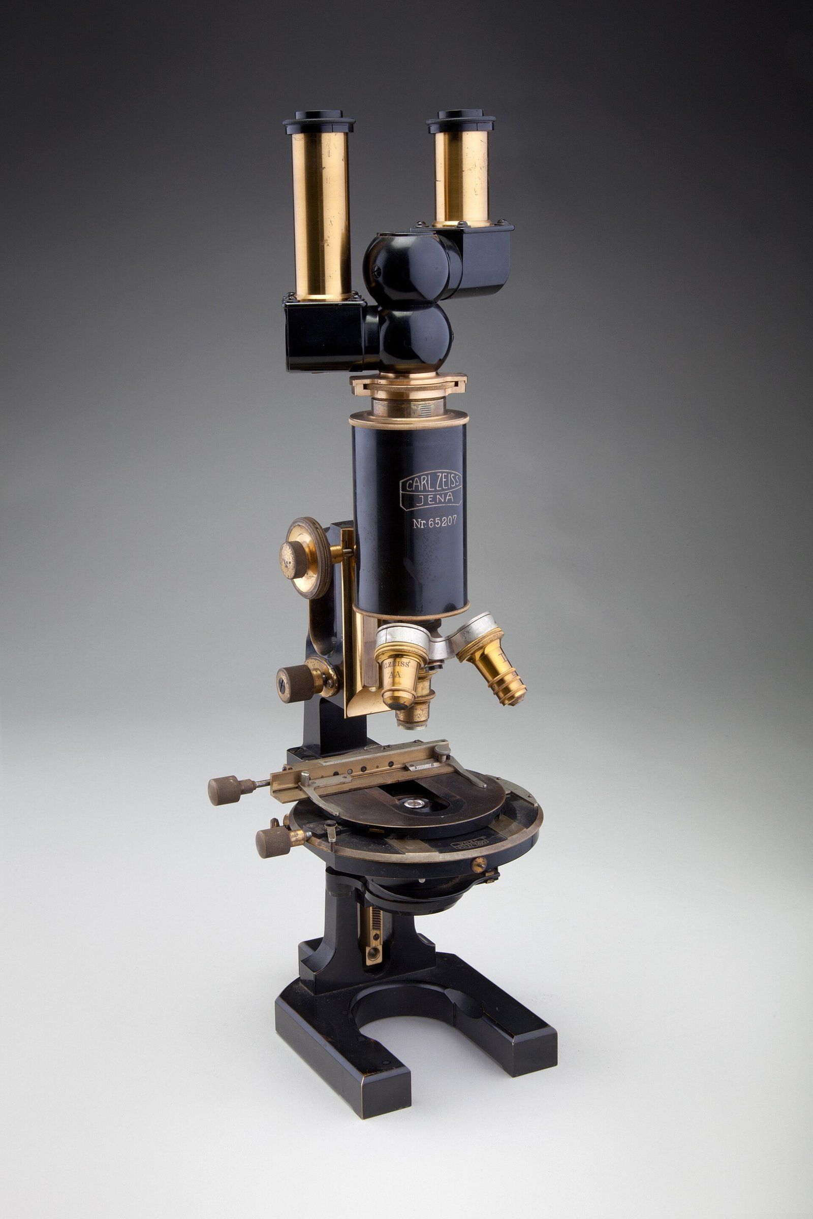
The Embryo Project Encyclopedia at Arizona State University records that the German gynecologist Hans Hinselmann built the colposcope in December 1924 in Hamburg and reported his findings in 1925, after years of searching for a better way to see the earliest changes of cervical cancer.4 The search began when his professor, Otto von Franqué, set him to study leukoplakia, white patches on the cervix that could be seen with the naked eye and that often lay beside small cancers.6 His device mounted a binocular microscope with a strong light on a stand. A 1967 TIME report described it as essentially a pair of binoculars with a light source on a pedestal, a principle that had barely changed, with lenses magnifying from six to 25 times and an examination lasting about seven minutes.9 Hinselmann expected to find tiny ulcers; O&G Magazine notes that he found instead a range of surface patterns reflecting changes in the tissue beneath.6
Stains and filters made changes visible
Two chemical aids shaped the method. In 1928 in Vienna, Walter Schiller introduced an iodine stain that marks abnormal cervical cells for biopsy, and in 1939 Helmut Kraatz described a green filter that sharpens the view of fine blood vessels.4 Hinselmann himself described the use of dilute acetic acid, essentially vinegar, in 1938, and its use spread quickly.6 Both the iodine and the acid remain part of the examination today.
How colposcopy took shape
- 1924Hans Hinselmann first uses his colposcope in Hamburg, reporting his findings the next year.
- 1928Walter Schiller introduces an iodine stain that marks abnormal areas of the cervix for biopsy.
- 1939Helmut Kraatz describes a green filter that sharpens the view of fine blood vessels.
- 1964Students of Karl Bolten found an American society for colposcopy.
- 1970sColposcopy becomes the standard follow up to an abnormal Pap test in the United States.
A slow road to routine use
The method spread slowly. A 2008 biography of Hinselmann on PubMed blames in part his insistence on his own complex terminology, his tie of the technique almost exclusively to early cancer detection, and the lack of teaching material until the 1950s.5 In the United States, early users found the instrument bulky, and the Pap test, developed at Cornell, became the main screening method by the 1960s.4 TIME reported that Karl Bolten, trained in Germany, arrived in 1952 and taught the method to scores of gynecologists.9 The Global Library of Women’s Medicine dates its entry into mainstream American gynecology to the late 1960s.8
A 2025 review marking the centenary describes colposcopy as now basic to the work of obstetricians and gynecologists worldwide.7
Why colposcopy is done
ACOG lists abnormal cervical screening results as the main reason, along with certain types of HPV infection. The examination can also look into vulvar itching or abnormal skin, genital warts, an inflamed cervix, polyps, pain or bleeding, and can check the result of earlier treatment.1 MedlinePlus adds that a Pap test can show abnormal cells but cannot provide a diagnosis; colposcopy gives the closer look that may confirm one. It may also follow a diagnosis of HPV, an abnormal area noticed during a routine pelvic examination, or bleeding after sex.2 The screening tests that lead to it are described in the history of the Pap test and in the entry on HPV and cervical cancer.
What the examination involves
The procedure is usually done in an office and takes 10 to 20 minutes, with about 10 more if a biopsy is taken.2 ACOG notes that it is best done outside a menstrual period, which gives a clearer view of the cervix.1 The colposcope stays outside the body while a speculum holds the vaginal walls apart, and a solution swabbed onto the cervix makes abnormal areas easier to see, sometimes with a slight sting.1 A biopsy may feel like a pinch or cramp, and cells may also be scraped from the canal of the cervix, a step called endocervical curettage. Afterward, spotting or a dark discharge for a few days is common, and after a biopsy there may be a day or two of discomfort. Heavy bleeding, severe lower abdominal pain, fever or chills are the warning signs ACOG lists.1
What happens during a colposcopy
- PositionThe patient lies on her back on an examination table with her feet in stirrups.
- SpeculumA speculum gently holds the vaginal walls apart so the cervix is visible.
- ColposcopeThe lighted, magnifying instrument stays outside the body, at the opening of the vagina.
- SolutionA vinegar or iodine solution is swabbed on, making abnormal tissue easier to see.
- SamplingAny area that looks abnormal may be biopsied, and cells may be taken from the cervical canal.
What the colposcopist looks for
The target is the transformation zone, the band where the cervix’s two kinds of lining meet. The Global Library of Women’s Medicine states that over 90 percent of cervical neoplasia arises there, and that the zone moves higher into the canal as a woman ages.8 After acetic acid is applied, abnormal tissue turns white; fine red dots, called punctation, or a tiled pattern, called mosaic, mark abnormal vessels, and sharp borders suggest a higher grade lesion. An examination counts as satisfactory only when the whole zone can be seen. Most instruments magnify between 2 and 40 times, and an iodine solution adds a second check: healthy tissue stains dark brown, while high grade lesions, poor in glycogen, do not.8
How the LEEP works
When a biopsy shows a higher grade change, the loop electrosurgical excision procedure removes the affected area of the cervix with an electrosurgical wire loop. A 2021 study on PubMed Central describes it as the preferred treatment for CIN 2 and CIN 3 lesions because it is safe, cost effective and practical under local anesthesia in an outpatient setting.3 It is also diagnostic, since it takes the whole transformation zone for examination under the microscope, and it is used when biopsy and cytology disagree or cancer is suspected.3 MedlinePlus notes that removing precancerous cells may keep them from developing into cancer, and that abnormal cells sometimes return.2 For that reason testing continues afterward. The Global Library of Women’s Medicine summarizes guideline options after treatment of CIN 2 or 3 as cytology at six and 12 months, cytology with colposcopy, or HPV testing, with colposcopy again if any result is abnormal.8
Biopsy and excision do not always agree
The same study compared biopsy results with the tissue later removed by LEEP in 241 women and found that the two matched in 41.9 percent of cases, in line with earlier reports of 40 to 57 percent. A biopsy graded low often proved milder still on excision, while a CIN 3 biopsy seldom did, and the authors called for methods that keep patients from being overtreated or undertreated.3
How often excision found a milder lesion than the biopsy
Timing matters too. Healio reported a study of 17,541 women with abnormal results in which those who had no colposcopy within a year carried about 2.3 times the later cancer risk of those examined within three months, while waiting three to 12 months made no measurable difference. The lead researcher suggested giving priority to high grade results and to women whose screening history is unknown.10
The range of normal
Many colposcopies find nothing serious. MedlinePlus lists genital warts, polyps and irritation of the cervix among possible findings, and says that a normal biopsy makes cells at risk of becoming cancer unlikely, though monitoring may continue because that can change.2 A normal cervix itself varies: in younger women the transformation zone often sits on the visible surface, while after menopause it may lie out of view within the canal. A red ring of glandular lining around the opening of the cervix, called ectropion, is a normal finding in younger women and becomes uncommon after menopause.8 What a given result means for one woman, and what follow up suits her, is for her own clinician to explain.
References
- American College of Obstetricians and Gynecologists. Colposcopy, November 2024.
- MedlinePlus. Colposcopy: MedlinePlus Medical Test.
- PubMed Central. The concordance between colposcopic biopsy and loop electrosurgical excision procedures in patients with known smear cytology and human papillomavirus results, 2021 12 31.
- Embryo Project Encyclopedia, Arizona State University. The Colposcope and Colposcopy (1925 to 1980), 2017 03 30.
- PubMed. History of colposcopy: a brief biography of Hinselmann, April 2008.
- O&G Magazine. Ab umbris ad lumina vitae: a short history of colposcopy, 2017 10 30.
- Journal of Obstetrics and Women's Diseases. History of the origin and development of colposcopy (dedicated to the 100th anniversary of colposcopy), 2025 07 23.
- Global Library of Women's Medicine. Colposcopy.
- TIME. Cancer: Direct Inspection, 1967 04 21.
- Healio. Cervical cancer risk rises with colposcopy delay after abnormal test results, 2023 10 03.
External links
See also
- Cervical Health and Screening
HPV and Cervical Cancer
A common virus, usually harmless, that lies behind almost all cancers of the cervix when a few types linger.
- Cervical Health and Screening
The HPV Vaccine
A shot given in early adolescence that is now measurably emptying the cervical cancer statistics of young women.
-
 Cervical Health and Screening
George Papanicolaou and the Pap Test
The Greek immigrant scientist, his guinea pig studies and the screening test that carries his name.
- Cervical Health and Screening
How Cervical Screening Guidelines Changed
From an annual smear to a virus test every five years: two decades of revised advice, and the reasons.
- The Gynecology Visit
The Well Woman Visit
A reference guide to the yearly preventive checkup for women, organized by age and reshaped by two decades of screening research.